Skin Cancer Diagnosis
Dermatoscopy
 A dermatoscope (aka a dermoscope) is a handheld device or a device connected to a larger unit which magnifies a skin lesion for more in depth examination. It utilizes polarized or non-polarized light to aid the examination and highlight different features elements not visible on naked eye examination alone.
Various methods and algorithms are used to assess the images. The current standard of practice is to capture these images and keep a digital record for future reference.
Biopsy of a Lesion
If a lesion is suspicious the only way to accurately diagnose it would be to do a biopsy. Many benign lesions can be mistaken for cancerous growths and vice versa. Even though our clinical and dermatoscopic exam can help distinguish between benign and malignant lesions, the most accurate way the confirm a diagnosis is by doing a biopsy and sending the specimen for histological analysis and assessment.
A biopsy can be excisional (the whole lesion is included with an appropriate border of adjacent skin) or incisional (part of the lesion is included). Choosing which method to use will depend on a few factors including the size of the lesion, the site (e.g. the earlobe versus the back) and the possible diagnosis. This will all be explained if and when a biopsy is planned.

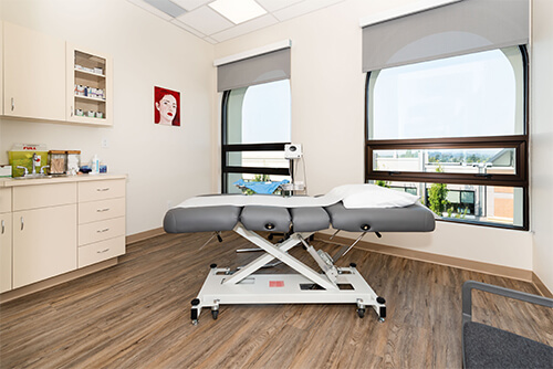
The biopsies themselves are sterile procedures and will be performed in our procedure room. We start by cleaning the site with an antiseptic and draping a sterile drape over the area. There after we anesthetize (“freeze”) the lesion and surrounding skin using Lidocaine (similar to dental “freezing”) and after a few minutes determine if the freezing was effective by testing the local reaction to pain stimulus. Bear in mind that there will be no pain or any feeling, but that deep pressure can still be perceived (we call this proprioception).
After we determined that the area is fully anesthetized, we will start to do the biopsy and remove the lesion (excisional) or part of it (incisional). Any small areas of localized bleeding (from for instance a small branch of an artery or vein) will be tied off (using dissolvable sutures) or cauterized (electrocautery, “burned”).
The final step would be to close the defect we caused and this could include deep dissolvable sutures (e.g. Monocryl or Vicryl) or just a primary skin closure. For the skin we use non-dissolvable sutures (e.g. Nylon) on the surface or dissolvable (e.g. Monocryl) subcuticular (just below the skin surface). The choice of skin closure can only really be determined the day of the procedure. Some biopsies (e.g. punch biopsies, a form of incisional biopsy) could be left to heal on their own.
After the procedure you will be provided with an information sheet to discuss wound healing and instructions for wound care.
After 5-14 days we prefer to see you back for wound assessment and if necessary removal of the sutures. If the Pathology results are available, we will discuss this also during the follow-up visit. If the results are not available, then we will contact you telephonically to inform you about the results and if a follow-up visit is necessary.
Other techniques in Skin Cancer Diagnosis
There are other methods which could aid the diagnosis of Skin Cancer but the majority of these methods are not readily available and still experimental.
- Confocal scanning laser microscopy (CSLM)
- Multispectral imaging
- Point-of-care non-invasive genetic testing
- Raman spectroscopy
